

What Are Cherry Angiomas?
Cherry angiomas are small vascular lesions that appear as red or purple bumps on the skin. They develop when clusters of tiny blood vessels form close to the surface of the skin.
They most commonly appear on the torso, arms, shoulders, and legs, although they can occur almost anywhere on the body. Cherry angiomas may develop individually or in groups and often become more common with age.
Because of their colour and appearance, they are sometimes referred to as red moles or cherry hemangiomas, although the terms angioma and hemangioma can refer to slightly different types of vascular growths.
Cherry angiomas are also known as Campbell de Morgan spots or senile angiomas and are very common in adults, with around half of people over the age of 30 developing at least one.

Are Cherry Angiomas Dangerous?
Cherry angiomas are completely benign, meaning they are not cancerous and do not usually require medical treatment.
They are harmless vascular growths that do not spread and are not associated with skin cancer. However, any new skin lesion can understandably cause concern.
Occasionally, cherry angiomas may be confused with other types of skin lesions, including pigmented moles or melanoma. For this reason, any growth that:
- Changes in colour
- Increases rapidly in size
- Bleeds without injury
- Develops an irregular shape
should be assessed by a qualified practitioner to confirm the diagnosis.
In most cases, cherry angiomas remain stable and cause no medical problems.
What Causes Cherry Angiomas?
The exact cause of cherry angiomas is not fully understood, but several factors are believed to contribute to their development.
Ageing
Cherry angiomas become increasingly common with age. Changes in the skin and blood vessels over time are thought to contribute to their formation.
Genetics
Some individuals appear more prone to developing cherry angiomas due to genetic factors.
Hormonal changes
Hormonal changes, including those that occur during pregnancy, may trigger the development of new angiomas or cherry hemangiomas.
Environmental factors
Certain chemical exposures and environmental factors have also been linked to the development of vascular skin lesions in some individuals.
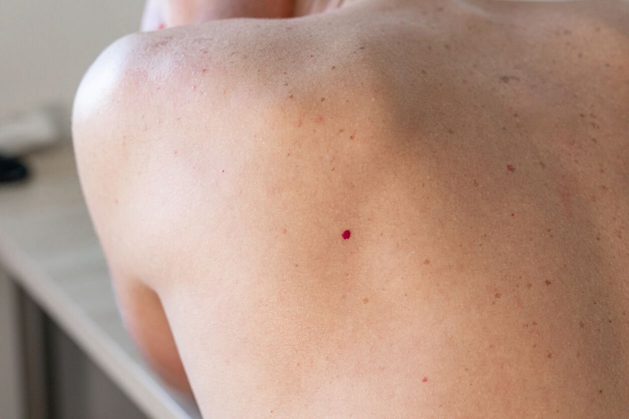
What Do Cherry Angiomas Look Like?
Cherry angiomas often begin as small, flat red skin spots and may gradually become slightly raised over time.
Typical characteristics include:
- Bright red, dark red, or purple colour
- Circular or oval shape
- Smooth or dome-shaped surface
- Small size, usually between 1 mm and 5 mm
They may appear on almost any body site, although they are most frequently seen on the chest, abdomen, back, arms, or shoulders.
Because they contain clusters of small blood vessels, cherry angiomas may bleed if scratched or irritated.
They are usually painless and do not cause itching or discomfort.

Can Cherry Angiomas Be Removed?
Cherry angiomas do not require treatment for medical reasons. However, many people choose to have them removed if they are:
- Cosmetically noticeable
- Increasing in number
- Frequently irritated by clothing
- Prone to bleeding after minor trauma
Removal should always be carried out by medically trained practitioners using appropriate dermatological skin treatments. Cherry angioma removal using laser therapy can safely target the blood vessels that form the lesion.
Attempting to remove cherry angiomas at home is not recommended, as this can lead to bleeding, infection, or scarring.
Laser Treatment for Cherry Angiomas
Laser and light-based treatments are commonly used to remove cherry angiomas by targeting the tiny blood vessels that form the lesion.
These advanced laser therapy technologies deliver controlled energy to the angioma, causing the blood vessels to coagulate and gradually fade while preserving the surrounding skin. This type of vascular laser treatment is designed specifically to target visible blood vessels.
At PHI Clinic, treatment may include advanced vascular laser therapy technologies such as Pulsed Dye Laser (PDL) or Intense Pulsed Light (IPL), depending on the characteristics of the angioma, the body site, and the patient’s skin type.
These approaches form part of a range of medically led treatment options designed to support long-term skin health while safely treating vascular lesions.
Pulsed Dye Laser (PDL)
Pulsed Dye Laser is designed specifically to treat vascular skin conditions. The laser emits pulses of light that are absorbed by the red pigment within blood vessels.
This energy helps collapse the tiny vessels that form the angioma, allowing the lesion to gradually fade over time.
PDL is particularly effective for:
- Small to medium cherry angiomas
- Red vascular lesions
- Angiomas on the chest, neck, arms, or face
Treatment is typically quick and minimally invasive, with little downtime.
Intense Pulsed Light (IPL)
Intense Pulsed Light uses a broad spectrum of light with specialised filters to target redness and visible vessels within the skin.
IPL may be recommended for patients with multiple small angiomas or when angiomas are present alongside diffuse redness, pigmentation, or sun damage.
In addition to reducing vascular lesions, IPL can help improve overall skin tone and clarity while supporting overall skin health.
Erbium YAG Laser
In selected cases, Erbium YAG laser may be used to precisely treat superficial skin lesions. This laser produces energy in the mid-infrared spectrum and allows controlled targeting of skin tissue while minimising impact on surrounding areas.
Treatment selection is always based on a detailed assessment to ensure the most appropriate approach for each individual and to identify the most suitable treatment options.
“Cherry angiomas are very common vascular lesions that tend to appear more frequently as we get older. While they are completely harmless, many patients prefer to remove them because they can become more noticeable on areas such as the chest, arms or face. With modern vascular laser technology we can treat them very precisely by targeting the tiny blood vessels that form the angioma, allowing it to fade without damaging the surrounding skin. For most patients the treatment is quick, straightforward and produces very satisfying results.”
What Results Can I Expect?
Most cherry angiomas respond well to laser treatment. Following treatment, the angioma may initially darken before gradually fading as the body clears the treated blood vessels.
Smaller lesions often improve quickly, while larger or multiple angiomas may require additional sessions.
Results vary depending on:
- The size of the angioma
- The number of lesions present
- Individual skin characteristics
Most patients experience minimal disruption to daily activities following treatment while maintaining healthy skin and overall skin health.
Is Cherry Angioma Treatment Safe?
When performed by experienced practitioners using medical-grade technology, laser treatment for cherry angiomas is considered safe and effective.
At PHI Clinic, all treatments begin with a detailed consultation. Practitioners assess skin type, lesion characteristics, and medical history to determine the most appropriate treatment approach.
Patient safety and skin health remain the priority at every stage of care. If you would like further information about cherry angiomas or available treatment options, our clinic support team can provide guidance and help you arrange a consultation.
“Cherry angiomas are completely benign but they can become more noticeable over time. Using targeted vascular laser technology we are able to treat the tiny blood vessels that create the angioma very precisely, allowing it to fade while preserving the surrounding skin.”
Safe and effective treatment
Your safety is our top priority and making our patients feel welcomed and reassured when entering PHI Clinic. To ensure maximum standard of patient care, all of our treatments are only ever carried out by our experienced and highly qualified team, who receive regular in-house and external training from industry experts.
At PHI Clinic we do not offer same day treatment as initial consultation to ensure patients have time to reflect on their bespoke treatment plan before choosing to proceed.

Cherry Angioma Frequently Asked Questions
Cherry angiomas are benign vascular lesions, meaning they are non-cancerous and harmless. However, any skin lesion that changes in size, shape, or colour should be assessed by a qualified practitioner.
Cherry angiomas are commonly associated with ageing. Changes in the skin and blood vessels over time can lead to the development of these small red lesions.
Yes. Because cherry angiomas contain small blood vessels, they may bleed if scratched, cut, or irritated.
Cherry angiomas typically do not disappear without treatment. They often remain stable over time, although more may develop with age.
Smaller angiomas may improve after one treatment, while larger or multiple lesions may require additional sessions. Your practitioner will advise during consultation.
Treated angiomas usually do not return, but new angiomas may develop elsewhere on the skin over time.
Most patients describe the sensation as mild and brief, similar to a quick snapping feeling on the skin. Treatments are generally well tolerated and quick to perform.
Location
PHI Clinic
102 Harley Street,
London,
W1G 7JB,
United Kingdom
Tel: 02070345999
Opening Hours
Monday & Tuesday - 9:30am to 6:00pm
Wednesday & Thursday - 9:30am to 8:00pm
Friday - 9:30am to 5:00pm
Saturday - 11:30am to 12:30pm
Sunday - Closed
Opening times can change, if your appointment is outside of these times please contact the clinic for confirmation.

